
Abstract:
Purpose
Spinal metastatic disease and multiple myeloma affects over 600,000 people every year in the United States and cause progressive bone destruction that results in incapacitating pain, fractures, and the inability to walk. Minimally invasive thermal therapies such as RF and ultrasound ablation techniques have proven to be effective for treating tumors outside the spine. The successes of such treatments rely on accurately controlling the direction and shape of the ablated region and avoid damaging anatomical sites such as the spinal cord. The purpose of the present study was to use a high intensity interstitial ultrasound ablator under ultrasound image and electromagnetic (EM) tracking guidance to ablate tissue near the lumber vertebrae in pig.
Methods
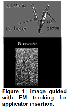
ExtensiveNeedle/catheter based interstitial ultrasound applicator of center frequency of 6-7 MHz was used to ablate 5 mm away from the dorsal process in the region at intersection of dorsal and transverse process in pig under gas anesthesia. Temperature sensors were placed at two different locations to estimate thermal distribution in the threedimensional treated volume. The ultrasound imaging was acquired using a SonixTouch (Ultrasonix, Richmond, BC, Canada) system with an L14- 5/38 GPS probe combined with EM tracking system to guide the insertion of the applicator (Fig. 1). An in-house software system for image processing and control was developed to communicate with the hardware, ultrasound imaging, tracking system, and treatment control.
Results
The ultrasound applicator was successfully inserted as per treatment plan using combined ultrasound imaging EM tracking guidance system. During treatment a maximum temperature of 60-70 °C and dose of 105-107 equivalent minutes at 43°C was observed at a distance 10 mm from the center of the ablation transducer for a treatment time of 5-7 minutes. The dose distribution was analyzed and compared with the gross pathology (Fig. 2) of the treated region. Conclusions: The experimental results demonstrated that the directionality and shape of the ablation zone can be controlled using the proposed technology of angularly-sectored tubular ultrasound transducers. This preliminary study shows the feasibility of ablating tissue near the vertebrae and in vertebral body successfully without damaging the spinal cord.
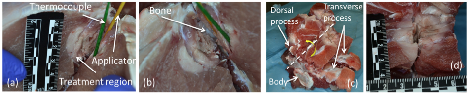
Figure 2: Gross pathology of the ablated muscle tissue near the spine with 360° pattern applicator showing (a) applicator, thermocouple position, and treatment zone, (b) the location of the bone and the ablated region, (c) anatomical sites of the spine and the treatment region (white dashed line indicate the line of cut for the tissue and yellow curved arrow indicate the directions tissue section was parted open to visualize the ablated region), and (d) a 3 cm by 3 cm cross-sectional area of the ablated region.